Flow Mediated Dilation
Flow Mediated Dilation or FMD is considered the Gold Standard in this field. However, the procedure is clinically challenging, requiring a high level of technical and human resource and skill. In the below article we discuss and propose how the FMD testing procedure and more so its evaluation can be facilitated.
Flow Mediated Slowing or FMS works according to the same basic principles as FMD, but can be carried out at a substantially lower entry level of cost and expertise, working mostly operator-independent.
While FMD and FMS address endothelial function of predominantly bigger vasculature, function tests of smaller vessels and the vascular bed are also available which may be advantageous in a normal population, e.g. in epidemiological studies and phenotyping.
Central to ’Gold Standard’ FMD testing is the availability of a high-resolution ultrasound scanner system. A linear array vascular probe of 7-12 MHz is employed for insonation of the brachial artery above the antecubital fossa. The diameter of the brachial artery, determined from the longitudinal ultrasound image, needs to be compared before and after brachial artery occlusion.
Although standards for the classical FMD exam have been published [6], testing procedures are quite demanding: The brachial artery needs to be insonated continuously at exactly the same position and angle for at least 8 to 10 min. The ECG triggered vessel diameter has to be determine manually or by a dedicated software at various points in time. Only experienced medical staff will be able to attain reproducible results.
-
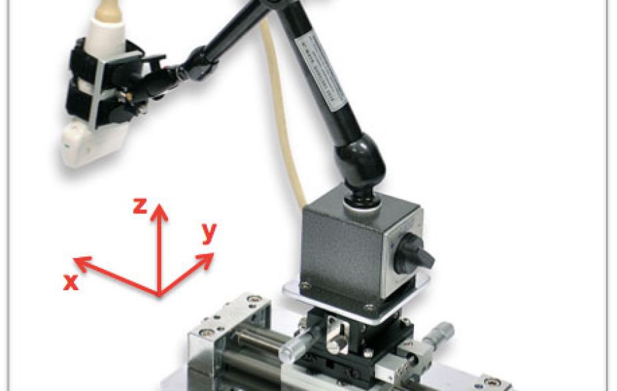
Probe Holder
 © Quipu.eu
Fig.: FMD Measurement - Probe Holder Prerequisite to the reproducibility of FMD, the employment of a micrometer adjustable probe holder is recommended for keeping the probe in exactly the same position throughout the test. This will also alleviate the operator from tiring probe holding, allowing more focus on the test subject, equipment adjustments, and image quality. A vacuum cushion should be available for stable and comfortable positioning of the arm.
-
FMD Evaluation
 © Quipu.eu
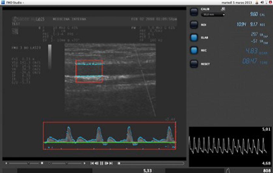
Fig.: Real-time evaluation of Flow Mediated Dilation test (by Quipu.eu, Cardiovascular Suite) For significant improvement of standardization, quality and reproducibility of FMD tests as well as for enhancement of operator-independence, a specialized real-time FMD evaluation software should be used. The figure shows an FMD test carried out with the validated Quipu Cardiovascular Suite employing a special algorithm for vessel diameter determination by continuous wall tracking [7]. The center window shows the image of the brachial artery, a real-time copy of the ultrasound scanner image on a PC. Marked in red, the area of interest evaluates the diameter of the carotid artery by a validated edge detector algorithm [8], abolishing the need of an ECG trigger. The software offers an automated evaluation procedure, independent of tedious and error prone manual diameter assessment. On the right side of the Doppler spectral display the instantaneous, pulsatile diameter variation is displayed. Below left, the mean diameter trace is shown The pre-occlusion rest period to the left (60s), the occlusion period (300 s) with a dark background, and the post-occlusive phase with the diameter increase and drop can be distinguished.
A comprehensive, thoroughly validated, and internationally widely adopted solution to FMD testing represents the Cardiovascular Suite offered by Quipu. Their system includes a proven micrometer probe holder, video grabber interfaces to virtually any ultrasound scanner, and the validated evaluation software on either Apple or Microsoft Windows PCs. Click here for more info and a free software trail. Watch the tutorial video of an automated FMD evaluation here.